 Why are they important?
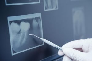
Without X-rays your dentist is limited to what he or she can see with the naked eye.
Many important problems can be missed without looking ‘beneath the surface’ with an X-ray, such as the early stages of decay between teeth which will lead to larger, more difficult and expensive to treat problems.
Dental X-rays are taken to:
- Find problems in the mouth such as tooth decay, damage to the bones supporting the teeth, and dental injuries (such as broken tooth roots). Dental X-rays are often done to find these problems early, before any symptoms are present.
- Find teeth that are not in the right place or do not break through the gum properly. Teeth that are too crowded to break through the gums are called impacted.
- Find cysts, solid growths (tumors), or abscesses.
- Check for the location of permanent teeth growing in the jaws of children who still have their primary (or baby) teeth.
- Plan treatment for large or extensive cavities, root canal treatment, placement of dental implants, and all tooth removals.
- Plan treatment for correcting crowded or badly aligned teeth (orthodontic treatment).
Why take different x-rays, isn’t one enough?
There are two main types of dental X-rays: intra oral (meaning the X-ray film is inside the mouth) and extra oral (meaning the X-ray film is outside the mouth).
- Intra oral X-rays are the most common type of dental X-ray taken.
These X-rays provide a lot of detail and allow your dentist to find cavities, check the health of the tooth root and bone surrounding the tooth, check the status of developing teeth, and monitor the general health of your teeth and jawbone.
- Extra oral X-rays show teeth, but their main focus is the jaw and skull.
These X-rays cover a wide area but do not provide the fine detail found with intra oral X-rays and therefore are not used for detecting cavities or for identifying problems with individual teeth.
Instead, extra oral X-rays are used to look for impacted teeth, cycsts, monitor growth and development of the jaws in relation to the teeth, and to identify potential problems between teeth and jaws and the temporomandibular joint (jaw joint) or other bones of the face.
Types of Intra oral X-Rays
There are several types of intra oral X-rays, each of which shows different aspects of teeth.
- Bite-wing X-rays show details of the upper and lower teeth in one area of the mouth. Each bite-wing shows a tooth from its crown to about the level of the supporting bone. Bite-wing X-rays are used to detect decay between teeth and changes in bone density caused by gum disease.They are also useful in determining the proper fit of a crown (or cast restoration) and the marginal integrity of fillings.
- Periapical X-rays show the whole tooth — from the crown to beyond the end of the root to where the tooth is anchored in the jaw. Each periapical X-ray shows this full tooth dimension and includes all the teeth in one portion of either the upper or lower jaw.Periapical X-rays are used to detect any abnormalities of the root structure and surrounding bone structure.
- Occlusal X-rays are larger and show full tooth development and placement. Each X-ray reveals the entire arch of teeth in either the upper or lower jaw.
Types of Extra oral X-Rays
There are several types of extra oral X-rays that your dentist may take.
- Panoramic X-rays (OPG) show the entire mouth area — all the teeth in both the upper and lower jaws — on a single X-ray. This type of X-ray is useful for detecting the position of fully emerged as well as emerging teeth, can identify impacted teeth, and aid in the diagnosis of tumors.
- Tomograms show a particular layer or “slice” of the mouth while blurring out all other layers. This type of X-ray is useful for examining structures that are difficult to clearly see — for instance, because other structures are in very close proximity to the structure to be viewed.
- Cephalometric projections show the entire side of the head. This type of X-ray is useful for examining the teeth in relation to the jaw and profile of the individual. Orthodontists use this type of X-ray to develop their treatment plans.
- Computed tomography, otherwise known as CT scanning, shows the body’s interior structures as a three-dimensional image. This type of X-ray, which may be performed in a hospital or radiology center rather than a dentist’s office, is used to identify problems in the bones of the face, such as tumors or fractures. CT scans are also used to evaluate bone for the placement of dental implants and difficult extractions. This helps the surgeon avoid possible complications during and after a surgical procedure.
Call us on (03) 5472 1377 or book your appointment online today.